Training
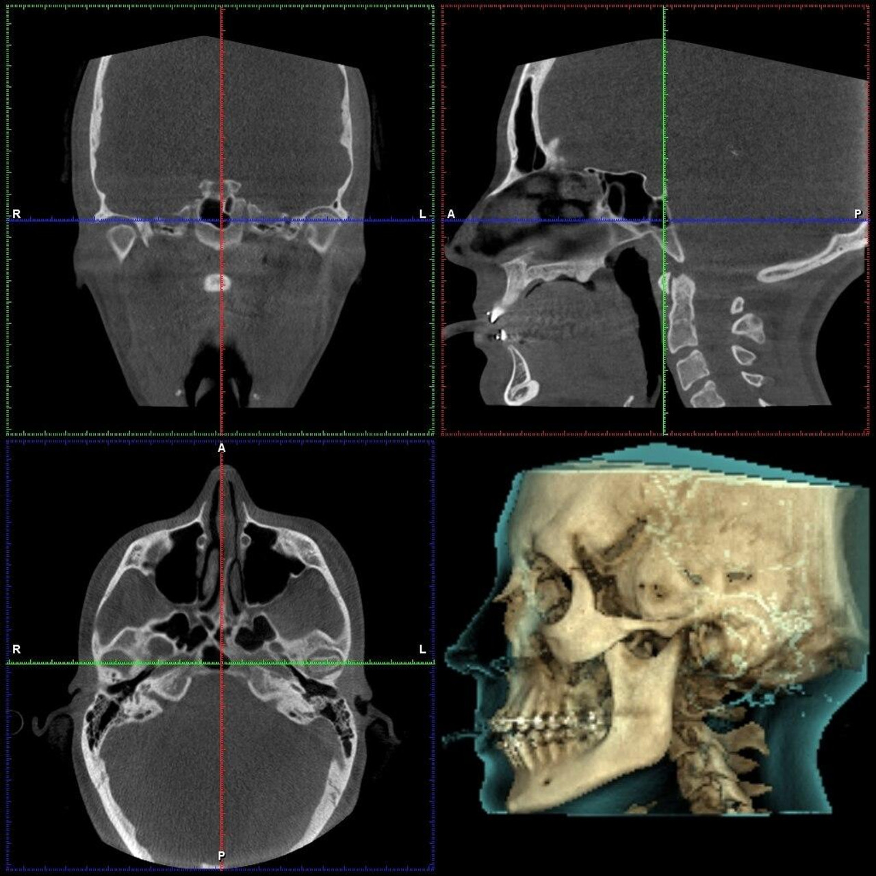
Cone beam computerized technology (CBCT) offers 3-dimensional visualization and more accurate imaging compared to analog and digital radiographs. This cutting-edge imaging technology enhances the dental practice. An adequate training and education program on its utilization and diagnostic-therapeutic potential while considering the risk benefit of ionizing radiation to patients proves prudent for an efficient dental practice.
This course will provide the practitioner with a thorough knowledge of the principles behind CBCT, including: CBCT image and technique selection; reducing radiation risk; understanding 3-D anatomical variations; recognizing key pathological processes; and the process of CBCT interpretation and reporting. The course content will follow the guidelines provided by European Academy of DentoMaxilloFacial Radiology 3D – Framework for Specialist Training in Dental and Maxillofacial Radiology 3D 2008.
Learning Objectives
- The course will provide an understanding of basic working principles of CBCT and radiation protection.
- Knowledge of the normal anatomical features seen on images taken of the teeth, oral cavity, jaws, facial structures and of the head and neck region.
- A deep insight into applications of CBCT scans in various realms of dentistry and beyond.
- Practical training sessions and hands-on case interpretations of volumes on CBCT software.
Basic
- What is CBCT?
- Fundamental principles and comparison with other imaging modalities.
- Setting up a CBCT Office
- Operational parameters and device settings
- Radiation Hazards and Dosimetry
- Knowledge and understanding of the relevant national regulations and guidelines of the country
- Anatomic review in various anatomic planes
- Artefacts on CBCT
- Interpretation of the images of disorders that affect the oral & maxillofacial region.
- Knowledge and understanding of the diseases that affect the oral & maxillofacial region.
- Introduction to the software followed by Hands-On.


Intermediate
- CBCT in Endodontics – Imaging protocols & interpretation of aberrant canal anatomy, localisation of accessory canals, calcified canals, root fractures, broken instruments, pulp stones etc.
- CBCT in pathologies of jaws and surgical planning
- CBCT in Pedodontics – considerations for a child patient
- CBCT in Periodontology – periodontal bone defects, bone grafting
- CBCT in Orthodontics, Impacted teeth and malocclusions
- Sleep disorders, airway analysis and TMJ analysis
- Image guided implant planning and assessment
Advance
- Digital Implant planning using digital impressions and CBCT scan on advanced implant planning software.
- Fabrication of surgical guides for implant placement.
- An insight into CBCT Sialography
